


Mapping flow of bio-electrical current to reveal brain’s inner workings
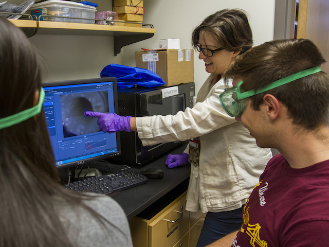
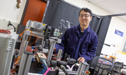
Rosalind Sadleir (middle) is leading research to develop methods of producing images of the most fundamental signatures of brain activity. Photo by Jessica Hochreiter/ASU.
Deeper understanding of the workings of the human brain will almost certainly open doors to significant medical advances.
Gaining that knowledge hinges to a large degree on finding new ways to peer closer into the brain’s basic mechanisms.
Arizona State University biomedical engineer Rosalind Sadleir is leading research to develop methods of producing images of the most fundamental signatures of brain activity.
“We want to directly image the cells that are involved and look at the detailed characteristics of these active brain structures,” said Sadleir, an assistant professor in the School of Biological and Health Systems Engineering, one of ASU’s Ira A. Fulton Schools of Engineering.
Employing her expertise in bio-electricity and neural engineering, she’s pursuing the goal by devising techniques to reveal the electrical properties of these cells.
“Electricity is the language of the body, so it’s important to pinpoint the active processes going on at the electrical level,” she explained
She is looking for action potentials – the hallmark of the processes that take place during the firing of a neuron. As that begins to happen, channels in neural membranes open, and ions move into and out of the cell.
The process causes a rapid increase in the voltage across nerve fiber. The electrical impulse can then spread down the fiber.
As an action potential happens, the conductance of a neural cell membrane changes, becoming around 40 times larger than it is when the cell is at rest. This changes the passage of an externally applied low-frequency electric current, and makes entire cells appear more conductive. Sadleir and her collaborators have recently demonstrated this in tiny samples of neural tissue taken from a sea slug, Aplysia Californica.
Sadleir’s lab team is conducting further measurements and validations of this observation using both sophisticated imaging and modeling approaches, with help from fellow faculty members in the School of Biological and Health Systems engineering, assistant professor Vikram Kodibagkar and research professor Mark Spano.
There also is a lot to be learned from mapping the broad distribution of conductivity in the whole head, Sadleir said.
By mapping the distribution of current – formed when an external electrical field is applied to the scalp – researchers hope to get a clearer idea of the mechanism underlying emerging therapies such as transcranial direct current stimulation, called tDCS. This therapy is being used for many applications, for example to affect cognitive performance, promote recovery after stroke and to treat epilepsy and depression.
“Because electrical currents take curved paths, and those paths are affected by the conductivity of the region they flow through, it’s difficult to accurately target electricity on a particular structure. Electrical current flows in a complicated way in the brain.” Sadleir said. “Our techniques allow us to actually measure and map the flow of tDCS currents in the brain.”
Being able to essentially watch the process happen can help researchers figure out why particular currents and electrode locations seem to have effects on certain brain functions “and help us understand how tDCS does what we think it does,” Sadleir said.
To accomplish this in her Neuro-Electricity Lab at ASU, she is using an advanced form of imaging known as magnetic resonance electrical impedance tomography (MREIT). The technology enables examining the behavior of single cells and detecting small changes in neural activity at the cellular level.
Her progress in these studies has earned support from the National Institutes of Health (NIH). One grant is to provide about $1.8 million over four years. A second NIH grant, through the National Institute for Neural Disorder and Stroke is providing about $410,000 over two years.
The imaging techniques she is trying to refine promise to give scientists and engineers one more effective way to seek answers to questions about how the brain operates.
In addition, her research findings could be applied to improving medical electrical-stimulation techniques used as therapeutic treatments for movement disorders – primarily Parkinson’s disease, but also epilepsy, muscular dysfunction, chronic pain and depression.
One project applies her imaging technique to studies to better understand the processes involved in perinatal brain injury in premature infants, using traditional electrical impedance tomography rather than magnetic resonance electrical impedance tomography. The work is being done in collaboration with the Oregon-based company Electrical Geodesics Inc., with support from an NIH Small Business Technology Transfer grant.
Media Contact
Joe Kullman, [email protected]
480-965-8122
Ira A. Fulton Schools of Engineering


